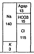
| Fig. 35. Serum electrolytes and the bar diagram | ||
|
| This section is part of Patil, Ramesh S. Causal Representation of Patient Illness for Electrolyte and Acid-Base Diagnosis. MIT Lab for Comp. Sci. TR-267 (1981). |
In this chapter we will consider in detail the two examples described in chapter 2. We will examine how the program accomplishes the tasks involved in these examples. Recall that the first example discusses a 40 year old 70 Kg male patient who has been suffering from moderately severe salmonellosis, and as a result, has developed moderately severe metabolic acidosis and hypokalemia. Recall also that the laboratory analysis of the patient's blood sample is:
| Fig. 35. Serum electrolytes and the bar diagram | ||
|
The program creates a top level PSD (the root node of the PSD tree) and instantiates the electrolytes in it. This PSD also corresponds to a PSM as it is the only PSD in the PSD tree. Next, the program generates possible acid-base disturbances that can account for the laboratory data. The acid-base analysis is based on the regression equations for the 95 per cent confidence intervals for acid-base disturbances [Schwartz65, Cohen66]. The nomogram of acid-base disturbances, the patient's acid-base state and the possible acid-base disturbances are shown in figure 36.
| Fig. 36. Graphical description of acid-base disturbances |
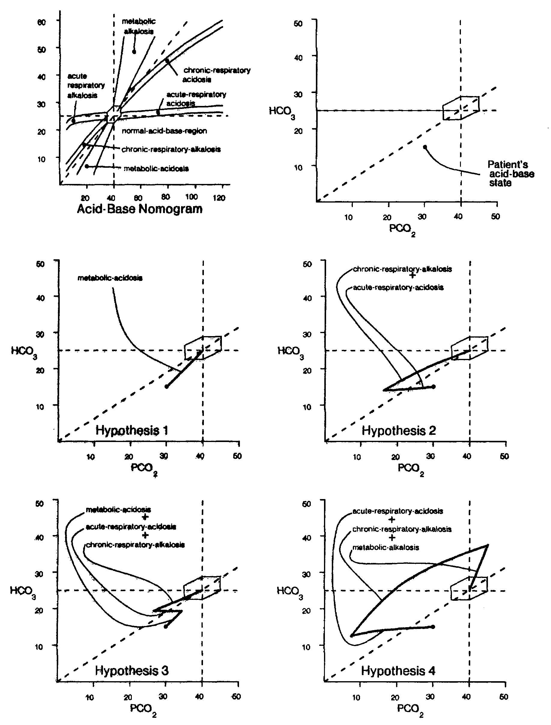 |
The list of these disturbances is rank-ordered and pruned. The rank-ordering is performed in two stages: first, by the complexity of the disturbance, and second, among the disturbances with same complexity by their seventies. For example, the complexity of the second acid-base disturbance is 2 (the number of components in the disturbance) and its severity is 0.75 = (0.682 + 0.322)0.5. The rank-ordered list is pruned to remove all the disturbances with more than two components from consideration during the initial formulation.28 The rank-ordered list of the likely disturbances is:
---- Patient Acid-Base Profile ----
1. metabolic-acidosis [sev: 0.4]                        very likely
2. chronic-respiratory-alkalosis [sev: 0.68]
   + acute-respiratory-acidosis [sev: 0.32]            unlikely
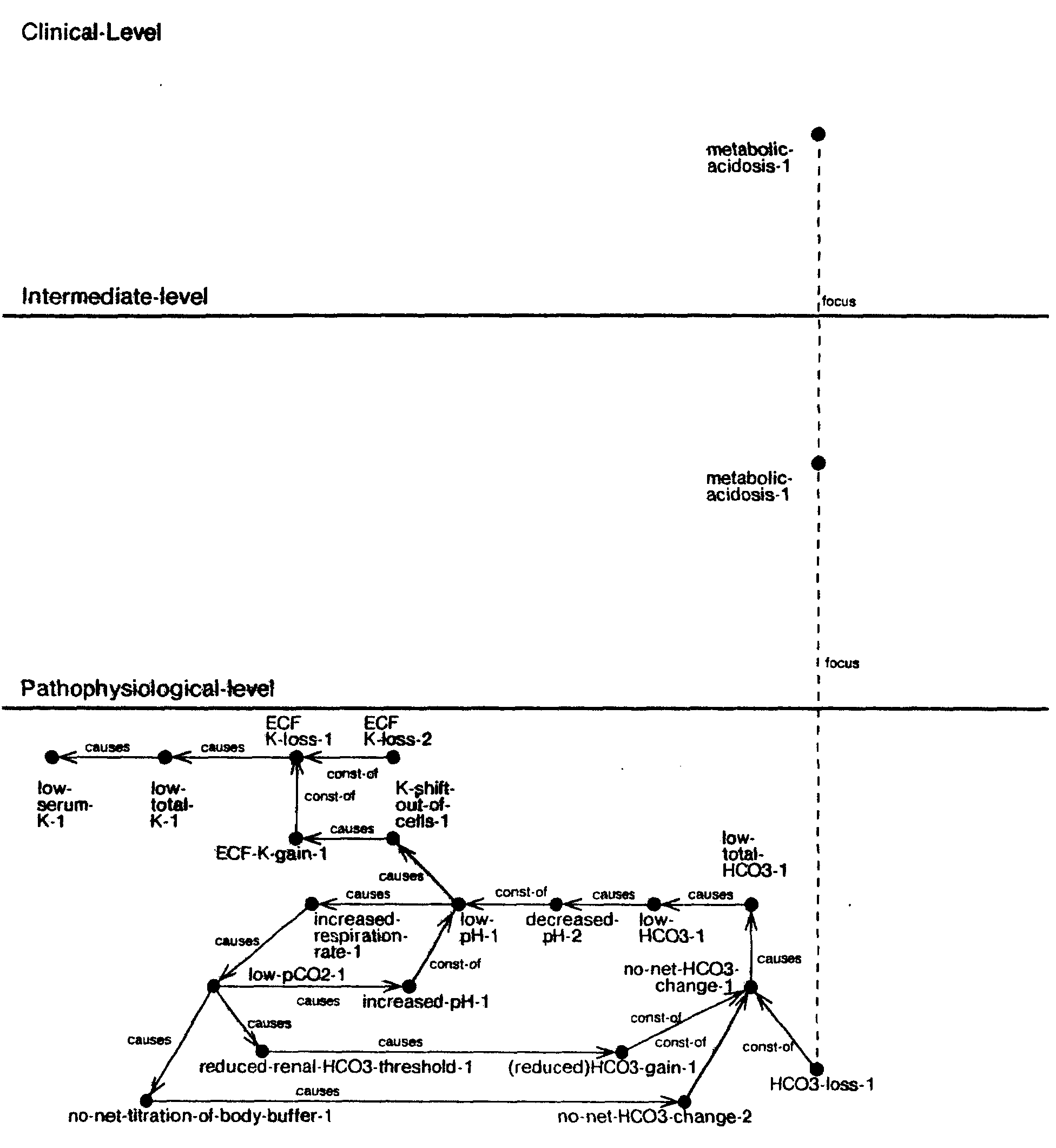
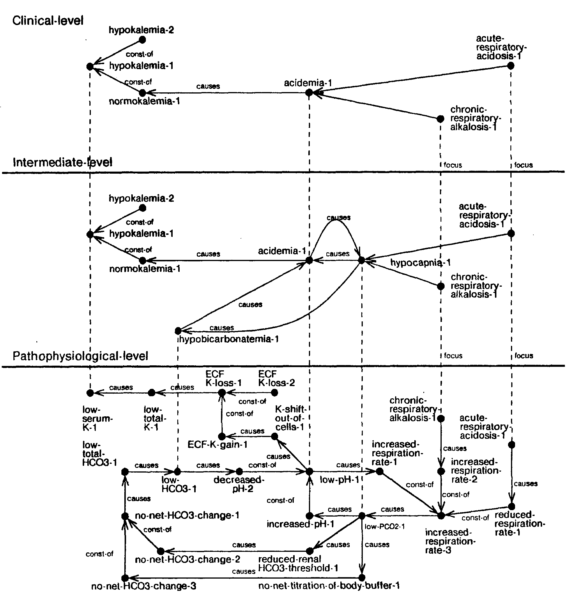
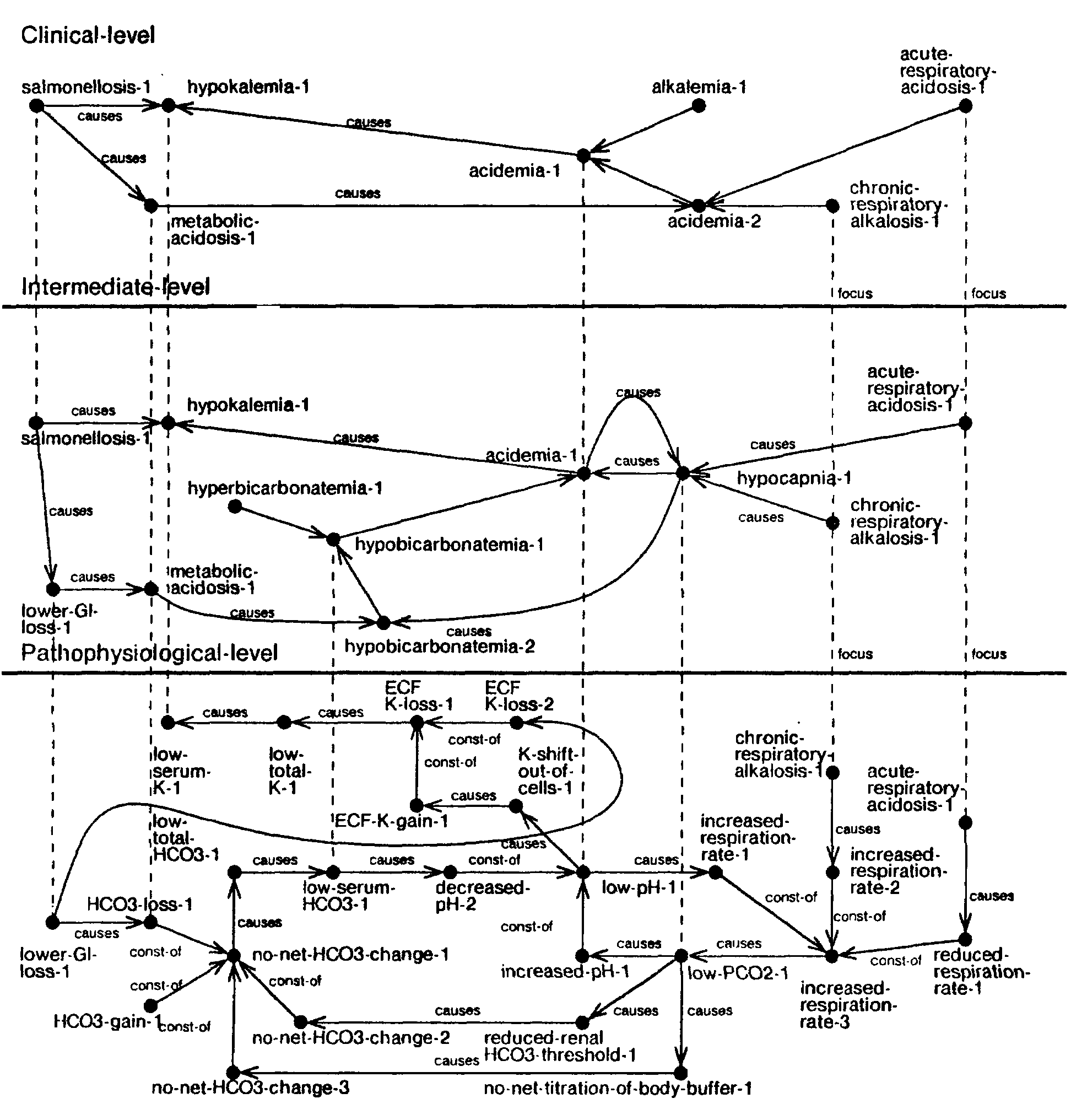
The two possible acid-base disturbances provide competing explanations of the serum electrolyte values. The program creates two inferior PSDs under the root PSD. It instantiates, at the clinical level, the nodes corresponding to metabolic-acidosis in the first, and chronic respiratory alkalosis and acute respiratory acidosis in the second (shown in figures 37 and 38). Next, it focally elaborates these nodes to the physiological level (the level at which the instances of electrolyte data are present). For example, in the first PSM the program focally elaborates the metabolic acidosis through the intermediate levels until it reaches the pathophysiological level and identifies the amount of HCO3 loss corresponding to the severity of the metabolic acidosis. Based on this information and the laboratory data, ABEL instantiates the feedback loop corresponding to the acid-base homeostatic mechanism. Next, it projects backward each node whose cause can be uniquely determined and projects forward the definite consequences of each node in the PSM.29 We now have the pathophysiological level explanation of the electrolyte abnormalities for each of the two likely acid-base disturbances (shown in figures 37 and 38).
| Fig. 37. Hypothesis 1 |
 |
| Fig. 38. Hypothesis 2 |
 |
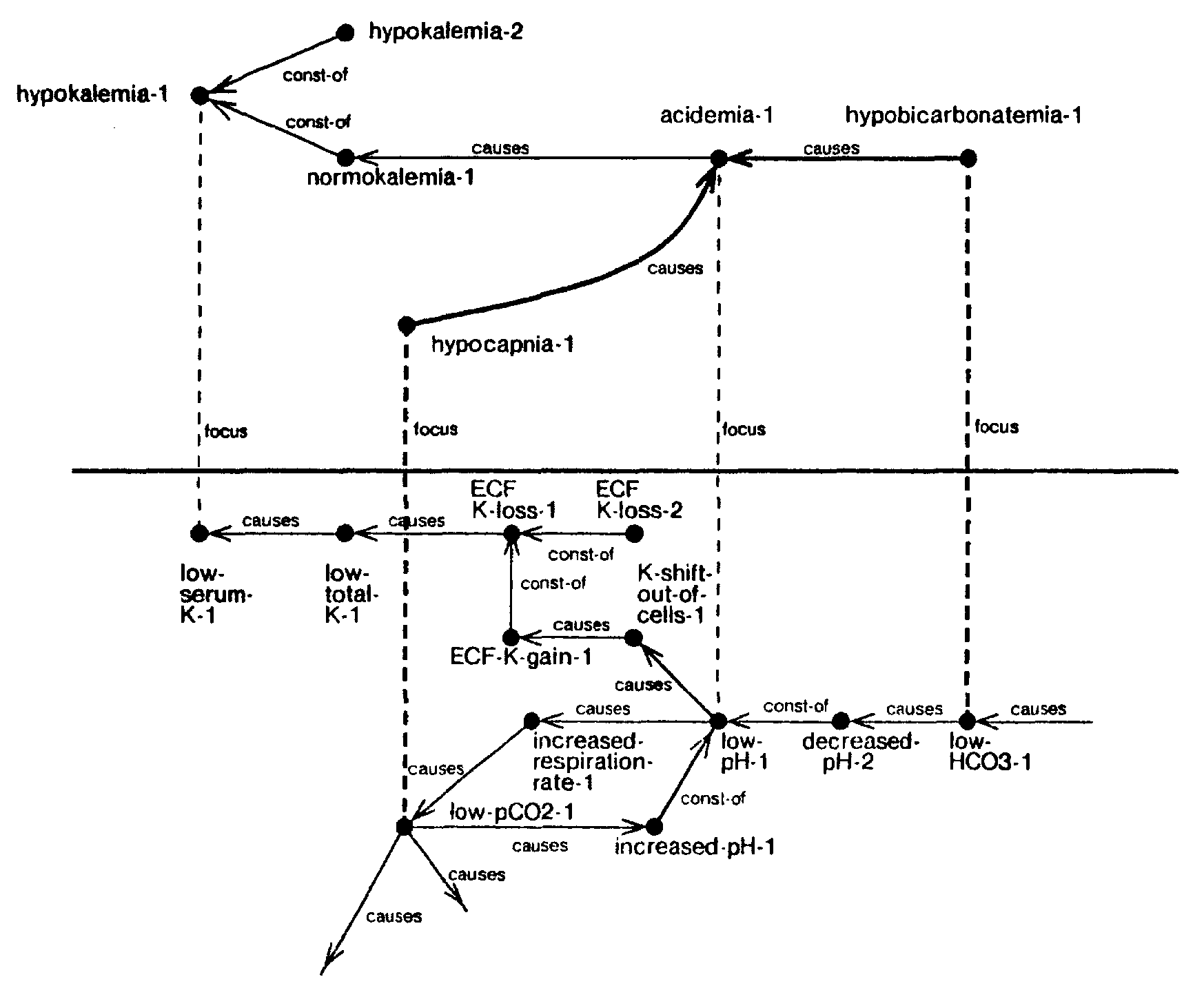
After the pathophysiological description is completed, it is aggregated, one level at a time, to the clinical level of detail. To illustrate this process let us consider the aggregation of the low- serum-K-1 node in PSM 1. Focally aggregating this node, we instantiate hypokalemia-1 as shown in figure 39. Next, we observe that one of the predecessor paths of low-serum-K-1 has an aggregable node on it, namely low-pH-1.30 We focally aggregate this node to instantiate one of the causes of hypokalemia. 1 (acidemia. 1) at the next higher level. Note that the other predecessor path from low-serum-K-1 does not have an aggregable node, therefore the component of low-serum-K-1 caused by this path must remain unaccounted for at the next higher level. Next, we compute the component of low-serum-K-1 that can be accounted for by low-pH-1 and the component that remains unaccounted because of the unaccounted ECF-K-loss-2. Then we compute the mapping of these two components at the next level of aggregation and instantiate normokalemia-1 (the component accounted for by low-pH-1) and hypokalemia-2 (due to unaccounted ECF-K-loss-2). We then causally connect the normokalemia-1 to acidemia-1 and mark the hypokalemia-2 as unaccounted. The structure added by the operations described above is shown in bold in figure 39.
| Fig. 39. Aggregation of low-serum-K-1 |
 |
| Fig. 40. Aggregation of low-pH-1 |
 |
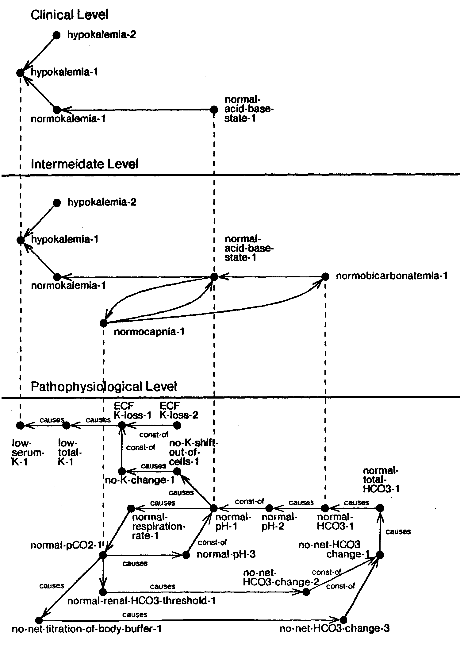
Next, let us consider the causal aggregation of low-pH-1 shown in figure 40. As each of the paths leading back from low-pH-1 has an aggregable node (low-pCO2-1 and low-HCO3-1),31 the focal aggregation of low-pH-1 (acidemia-1) is a fully accounted node. The causal aggregation is achieved by focally aggregating the low-pCO2-1 and low-HCO3-1 into hypocapnia-1 and hypobicarbonatemia-1 respectively, and by causally connecting hypocapnia-1 and hypobicarbonatemia-1 to acidemia-1. This process is repeated for each aggregable node at the current (pathophysiological) level and then the whole process is repeated at the next level until we reach the clinical level of aggregation. The resulting structures for the two acid-base hypotheses (encoded by the two PSMs) are shown in figures 41 and 42.
| Fig. 41. PSM for hypothesis 1 |
 |
| Fig. 42. PSM for hypothesis 2 |
 |
As discussed in chapter 2, a comparison of the clinical level explanations shows that the two PSMs share the structure involving hypokalemia and acidemia. They differ in their accounting for acidemia. Note that the acid-base feedback cycles present at the pathophysiological and intermediate levels have been abstracted away by the aggregation process and the two clinical level descriptions are fairly simple. A comparison of the intermediate level descriptions shows that they differ principally in the way the acid-base feedback cycle is perturbed. In the first case, the change in acid-base state is a consequence of addition of H+ to the body which causes hypobicarbonatemia, whereas in the second it enters as primary disturbance in ventilation which alters the CO2 tension. The pathophysiological level differences between the two cases can be identified similarly by comparing the two pathophysiological level descriptions. Finally, note that the first PSM has two unaccounted findings while the second PSM has three unaccounted findings.
In the context of this initial analysis the program starts its diagnostic exploration. It computes the diagnostic closures for the two hypotheses (DC-1 and DC-2 shown in figures 43 and 44), and formulates the top level goal of pursuing DC-1. One complete cycle of diagnostic inquiry is shown in figure 45.
| Fig. 43. Diagnostic closure 1 |
 |
| Fig. 44. Diagnostic closure 2 |
 |
As a first step towards exploring DC-1, the program groups the disease hypotheses according to the number of unexplained findings each disease hypothesis can explain. For example, the salmonellosis hypothesized to account for moderately severe metabolic acidosis can also account for the hypokalemia. Therefore, the hypothesized salmonellosis can account for all the unaccounted findings in PSM-1. However, if the patient had very severe metabolic acidosis and mild hypokalemia, the salmonellosis hypothesized to account for metabolic acidosis would not have been consistent with hypokalemia. In such a case we would have had to hypothesize two separate instances of salmonellosis, each accounting for only one of the two unaccounted findings. Subsequently, each of the two instances of salmonellosis would have been grouped with disease hypotheses accounting for only one unaccounted finding.
| Fig. 45. One complete cycle of diagnostic inquiry |
Differentiating between the causes of                          (a)
the leading complete hypothesis.
     1 SALMONELLOSIS
     2 URETEROSIGMOIDOSTOMY
     3 VILLOUS-ADENOMA
     ------------------------
     4 DISTAL-RTA
     5 PROXIMAL-RTA
     6 ACUTE-RENAL-FAILURE
     7 CHRONIC-RENAL-FAILURE
continue? ==> y
Does the patient have any of the following?                    (b)
     1 SALMONELLOSIS
     2 URETEROSIGMOIDOSTOMY
     3 VILLOUS-ADENOMA
Present: ==> none    Absent: ==> none    Unknown: 1 2 3
I would like to ask about the effects of SALMONELLOSIS. Does the patient have one of the following? (c) 1 DEHYDRATION 2 EDEMA Present: ==> none Absent: ==> none Unknown: 1 2 Is the value of SERUM-CREATININE known? ==> yes (d) Please enter the attributes of SERUM-CREATININE
What is the VALUE of SERUM-CREATININE ? ==> 3
What is the START-TIME of SERUM-CREATININE ? ==> 0
Is the value of MEAN-ARTERIAL-BLOOD-PRESSURE known? ==> yes (e) Please enter the attributes of MEAN-ARTERIAL-BLOOD-PRESSURE What is the value of MEAN-ARTERIAL-BLOOD-PRESSURE ? ==> 75 |
Next, the diseases in the same group are rank-ordered based on their scores computed from the three factors, match, mismatch and unknown (described in section 5.4). Those hypotheses which have higher mismatch than match are not considered. For example, consider the scoring of the vomiting hypothesized to account for unaccounted hypokalemia. The vomiting so hypothesized matches the hypokalemia. However, the hypothesized vomiting predicts metabolic- alkalosis which is inconsistent with the observed metabolic acidosis. Furthermore, if vomiting were really observed in the patient, the additional amount of HCO3 loss necessary to cause the observed state would require a very severe cause of metabolic acidosis to be present. Therefore, vomiting has a substantially higher mismatch factor as compared to the match and it is rejected. The program deletes the hypotheses that have been rejected and rank-orders the remaining as shown in figure 45(a).
Based on the categorization of the disease hypotheses, ABEL decomposes the diagnostic problem into two groups by constructing two separate diagnostic closures (DC-3 and DC-4). DC-3 (shown in figure 46) contains disease hypotheses 1 to 3, and DC-4 contains disease hypotheses 4 to 7. It projects forward the disease hypotheses in each of the two diagnostic closures to identify their unobserved findings. Next, it sets up a goal to differentiate between the three hypotheses in DC-3. As the first step towards this differentiation, the program asks if the user is already aware of any of the possible alternatives as shown in figure 45(b).
| Fig. 46. Diagnostic closure 3 |
 |
When none of the three hypotheses can be directly confirmed, the program pursues the task of differentiating between the three further. It sets up an individual diagnostic closure for each of the three alternatives (DC-5, DC-6, and DC-7) and selects the next item (dehydration) for inquiry.32 Note that salmonellosis, ureterosigmoidostomy and villous-adenoma all cause dehydration. However, the program also notices that some of the diseases in DC-4 (e.g.., chronic-renal-failure) may have the exact opposite effect of causing edema. Therefore, while exploring dehydration (state of extracellular fluid volume) the program includes edema in the question (shown in figure 45(c)). The program is expecting dehydration. Therefore, when it fails to confirm or deny the dehydration it pursues the finding further (figure 45 (d) and (e)).
| Fig. 47. Diagnostic closure 4 |
 |
| Fig. 48. Diagnostic closure 5 |
 |
The program has now completed one full cycle of its diagnostic inquiry. It incorporates the information gained during this cycle in both the PSMs. Note that the program has already gathered sufficient information to confirm salmonellosis. It is unable to do so because we have not implemented the criteria for confirming a disease yet.33 Therefore, the program starts the new cycle of diagnostic planning in which it attempts to rule out all other possible causes of the acid-base disturbance. Finally, when it has exhausted all the findings relevant to the diagnosis of this case, it concludes that salmonellosis is the leading candidate and asks if the user would like to assume salmonellosis (shown in figure 49).
| Fig. 49. After all findings have been exhausted |
All possible etiologies that could explain the patient's
illness are unknown. In order to proceed we must at least
hypothetically assume one of them. Possible etiologies that could
explain the patient's illness listed in decreasing order are:
1 SALMONELLOSIS ---------------------------- 2 VILLOUS-ADENOMA 3 URETEROSIGMOIDOSTOMY ---------------------------- ---------------------------- 4 DIABETES-INSIPIDUS ---------------------------- 5 DISTAL-RTA 6 PROXIMAL-RTA Would you like to assume SALMONELLOSIS ? ==> yes Assuming MODERATE ACUTE SALMONELLOSIS. |
The program adds salmonellosis to the patient models and re-evaluates the two hypotheses. The process of assimilating salmonellosis into the PSMs is described next. Let us first consider the operation of causally connecting salmonellosis with metabolic-acidosis in PSM-1. As the observed salmonellosis is consistent with the metabolic acidosis, a causal link from salmonellosis to metabolic acidosis is established at the clinical level. The elaboration operator is used to establish this relation at the more detailed levels (the resulting structure is shown in figure 50).
| Fig. 50. Hypothesis 1 with salmonellosis |
 |
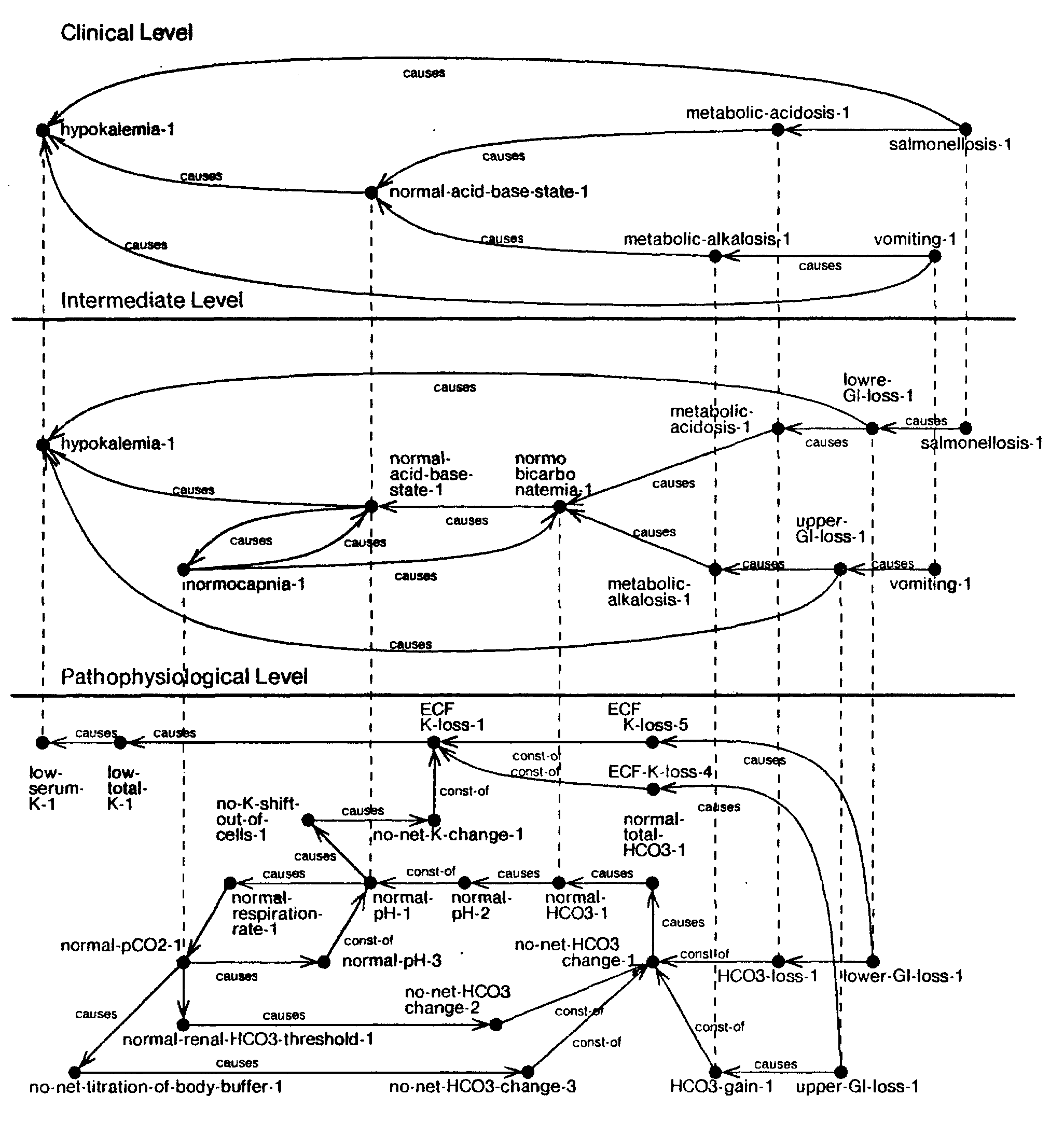
The elaboration process begins with the focal elaboration of salmonellosis to the intermediate level. The focus nodes of the source and the destination of the link being elaborated (salmonellosis —causes—> metabolic-acidosis) are now present at the next level. Next, ABEL attempts to match the causal path associated with the link at the next level of detail, namely salmonellosis —causes—> lower-GI-loss —causes—> metabolic-acidosis. As this path does not exist at the intermediate level, ABEL must establish this path and then proceed to elaborate each link in it. Let us first consider the link salmonellosis-1 —causes—> lower-GI-loss-1. As salmonellosis is a primitive node at this level (it does not have a focal node at the next lower level), the link between salmonellosis and lower-GI-loss is a primitive link and cannot be elaborated any further. The next link, lower-GI-loss-1 —causes—> metabolic-acidosis-1 however, can be elaborated further. This is done by first focally elaborating lower-GI-loss-1 at the intermediate level to the pathophysiological level, and second by connecting, at the pathophysiological level, lower-GI-loss-1 to HCO3-loss-1. As the remaining links in the causal path at this level are already present, this completes the process of elaboration. Next, the newly created structure is causally aggregated to propagate the consequences of the lower level additions back up to the clinical level. The results of assimilating salmonellosis into the two PSMs are shown in figures 50 and 51.
| Fig. 51. Hypothesis 2 with salmonellosis |
 |
A comparison of PSM-1 and PSM-2 shows that PSM-1 contains only one acid-base disturbance, while PSM-2 contains three acid-base disturbances. All the findings in PSM-1 have been accounted for, while PSM-2 has three nodes that still need to be accounted for. Therefore, based on the assumption that the patient is suffering from moderately severe salmonellosis, ABEL concludes that PSM-1 provides an adequate explanation of the patient's illness. The computer generated English descriptions of the clinical levels of the two PSMs are shown in figure 52.
| Fig. 52. English description of the two hypotheses | ||||
|
The second example deals with a patient suffering from moderately severe vomiting and salmonellosis. Recall that the electrolyte and acid-base disturbance in vomiting results from an excessive loss of upper gastrointestinal fluid, whereas in salmonellosis it results from an excessive loss of lower gastrointestinal fluid. The upper GI fluid is acidic while the lower GI fluid is alkaline, therefore the two tend to have offsetting effects on the acid-base balance. However, vomiting and salmonellosis both cause hypokalemia and volume depletion, therefore they compound the effects of each other.
In this example, we will consider the presentations of vomiting and salmonellosis such that they exactly offset the acid-base effects of each other, leaving the patient with no net change in pH. We will demonstrate the program's capabilities in dealing with multiple etiologies and in reformulating its patient description when new information is provided. We will illustrate this by describing the program's understanding of the case at three points during the diagnostic process: (1) just after the initial presentation of electrolytes, (2) after the program has identified the first of the two diseases, namely vomiting, and (3) at the end of the diagnostic process.
The program's initial evaluation of the patient's electrolytes is as follows:
---- Patient Acid-Base Profile ----
   1. normal-acid-base-state
This is a 40 year old 70 Kg male patient with moderate hypokalemia. His electrolytes are: Na: 143.0 HCO3: 25.0 Anion Gap: 12.0 K: 2.0 PCO2: 39.0 Cl: 108.0 pH: 7.42The hypokalemia remains to be accounted for.
The initial PSM (PSM-1) created by the program is shown in figure 53. Note that the clinical level of the PSM contains only one abnormal finding, hypokalemia. Figure 54 shows the revised PSM after vomiting has been introduced. A detailed description of this process of revision is considered next.
| Fig. 53. Initial PSM |
 |
| Fig. 54. Revised PSM after vomiting is entered |
 |
Based on the information in the diagnostic closure the program concludes that the vomiting present cannot account fully for the hypokalemia. However, as the vomiting can partially account for the hypokalemia (leaving a smaller amount unaccounted for), the program decides to project forward, to identify the quantity of hypokalemia accounted for by it. The projection process begins with the focal elaboration of vomiting-1 from the clinical level to the intermediate level. Next, the program matches the causal path associated with the link, i.e., vomiting —causes—> upper-GI-loss —causes—> hypokalemia. As this path is not inconsistent with the PSM, the program recurs on each link in the path. The first link, vomiting —causes—> upper-GI-loss, is a primitive link. Therefore, the program instantiates the upper-GI-loss (upper-GI-loss-1) and the link connecting it upwards to vomiting.1. The second link, upper-GI-loss —causes—> hypokalemia, is a compound link. The path associated with this link at the next level is upper-G I-loss —causes—> ECF-K-loss —causes—> low-total-ecf-K —causes—> low-serum-K. Matching this path with the description in the PSM, the program finds that all but one link, upper-GI-loss —causes—> ECF-K-loss, is already present. Since this link is primitive, the program revises the component structure of ECF-K-loss-1 and instantiates the link between ECF-K-loss-4 and upper-Gl-loss-1. Note that as soon as this link is instantiated the path at the pathophysiological level is complete. The program aggregates back the effects of the projection process to reflect the additions at the lower levels at the upper levels.
An important side-effect occurs when the program is reasoning (at the pathophysiological level) about the quantity of ECF-K-loss associated with the upper-GI-loss. As the ECF-K-loss is dependent on the quantity of upper gastrointestinal fluid loss, this loss must be accompanied by the loss of corresponding amounts of the other electrolytes present in the upper-GI-fluid, notably the loss of H+.34 This fact is incorporated into the PSM, causing the program to revise its acid- base hypothesis. This hypothesis now contains two components: an alkalemia (metabolic-alkalosis) caused by vomiting, and an acidemia (unaccounted) which cancels the effects of alkalemia leaving the patient in a normal acid-base state as shown in figure 54. Thus, the PSM after vomiting contains two unaccounted nodes: the unaccounted component of hypokalemia (less severe than before vomiting was introduced), and acidemia which must be present to offset the metabolic-alkalosis caused by vomiting.
Note that the two unaccounted components of the PSM are the same as those present in PSM-1 of the first example. We have been successful in separating the effects of vomiting from the remaining disturbance (salmonellosis). As might be expected, from here on the diagnosis of this case is similar to that of the first example. The final diagnosis after salmonellosis has been added to the PSM is shown in figure 55. Figure 56 shows the program's English explanation of the final diagnosis at two different levels of detail.
| Fig. 55. Final PSM after salmonellosis is introduced |
 |
| Fig. 56. English text of the final explanation | ||||
|
This section is part of
The document was reconstructed for the Web in April 2002 by Peter Szolovits.Patil, Ramesh S. Causal Representation of Patient Illness for Electrolyte and Acid-Base Diagnosis. MIT Lab for Computer Science Technical Report TR-267. October 1981. Also: Ph.D. Thesis, MIT Dept. of Electrical Engineering and Computer Science.