


Next: Stain cells to enable
Up: Quantitate expression levels of
Previous: Quantitate expression levels of

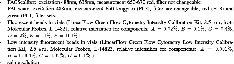
Materials:
Methods:
- Keep fluorescent beads refrigerated in
 C before use.
Protect from light. Do not freeze.
C before use.
Protect from light. Do not freeze.
- Set up FACScan to the following parameters: ?????????
- Vortex mix the vial contents to ensure that the polystryrene
beads are uniformly suspended.
- Add one drop from each vial to 2 mL of Haema-Line sheath fluid
or buffered saline solution. Notice that vial components C and D
from the low intensity kit are the same as vial components A and B from
the normal intensity kit.
- Sonicate the diluted bead suspension to disperse aggregates and
vortex mix immediately before use.
- Load unto FACScan, run for 10-60 seconds, then start recording
data.
- For each different fluorescent bead, should see a sharply
defined peak. Experimental data:
- Store bead fluorescense data for use in calibrating GFP
fluorescense from live samples.
Ron Weiss
Wed Feb 10 15:48:08 EST 1999